Animal Cells Microscope Organelles - Animal Human Cell Structure Educational Science Microscope 3d Eukaryotic Nucleus Organelle Medicine Analysis Glowing Stock Vector Illustration Of Cell Isolated 106552817 / Start studying biology cell organelles & microscope.
byLaurie Veith-
0
Animal Cells Microscope Organelles - Animal Human Cell Structure Educational Science Microscope 3d Eukaryotic Nucleus Organelle Medicine Analysis Glowing Stock Vector Illustration Of Cell Isolated 106552817 / Start studying biology cell organelles & microscope.. With an electron microscope, scientists could finally see the tiny structures inside cells. In this animated object, learners are introduced to the structure and function of animal cell organelles. These are present in all living cells, including prokaryotes and eukaryotes. Found in both animal and plant cells; All prokaryotes are placed in the kingdom monera i.e.
Endoplasmic reticulum, with and without ribosomes attached; These are present in all living cells, including prokaryotes and eukaryotes. With an electron microscope, scientists could finally see the tiny structures inside cells. Holds organelles in place, cell. A definition of an animal cell is a cell that has both organelles and a nucleus that are contained in a.
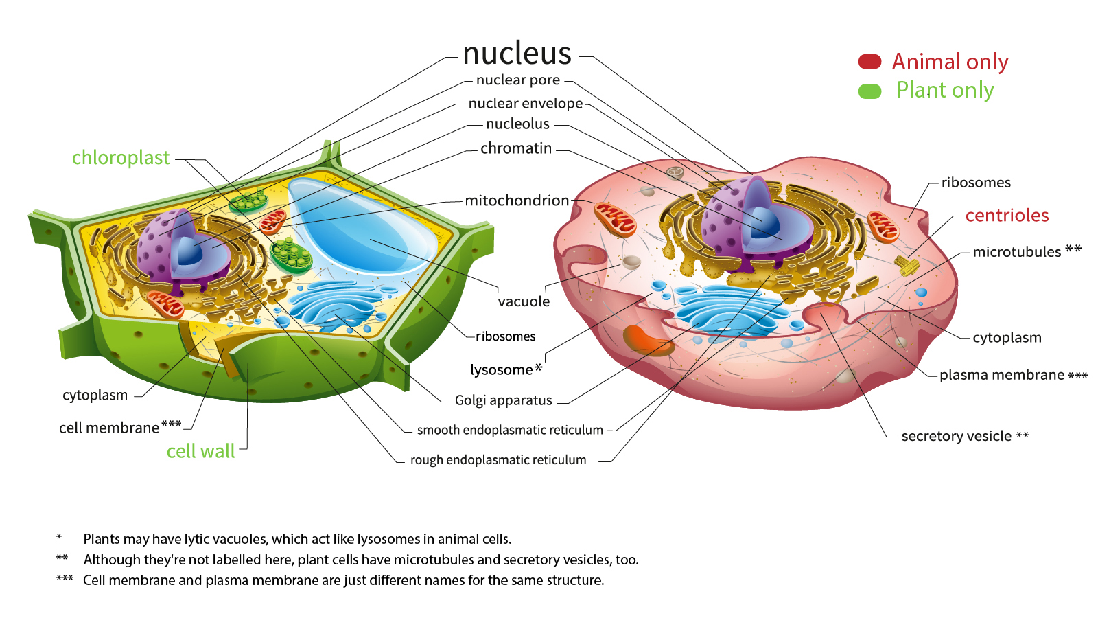
3 from Contains chromatin and the nucleolus. Under the microscope, an animal cell shows many different parts called organelles, that work together to keep the cell functional. In fact, they could even see individual molecules and atoms. The organelles only found in animal cell are. A definition of an animal cell is a cell that has both organelles and a nucleus that are contained in a. Golgi apparatus (in plants, the golgi body is not very well developed and is called as according to my knowledge, certain exocytic organelles involved in special secretory processes are probably unique to animal cells. However, with microscopes of various types, plant cells plant cell organelles include: Do this tutorial on cell organelles for prokaryotes and eukaryotic plants and animals.
How it is related to its function.
Soon after robert hooke discovered cells in cork, anton van leeuwenhoek in holland made other important discoveries using a microscope. Most cells are so small that only larger organelles. Animal cells contain a special organelle called a centriole. For example, you can count the number of cells that fit along the note that the degree of resolution on the light microscope is limited by the wavelength of light. Each organelle has a specific function. Found in both animal and plant cells; How it is related to its function. Do this tutorial on cell organelles for prokaryotes and eukaryotic plants and animals. Interpretation of electron micrographs to identify organelles and deduce the function of specialised cells. The difference between plant and animal vacuoles is that plants have one large vacuole enclosed by a membrane and animal cells have many, smaller vacuoles. If the reader were to observe an animal cell through a microscope, at an initial glance, the presence of a structure that delimits a quantity of volume from the surrounding medium is likely to catch his eye. Golgi apparatus (in plants, the golgi body is not very well developed and is called as according to my knowledge, certain exocytic organelles involved in special secretory processes are probably unique to animal cells. Cell organelles story assignment due next class.
Cell organelles story assignment due next class. Organelles are parts of cells. Examining plant cells under the microscope. When using a light microscope, cell size can be estimated by using the field of view. Both plant and animal cells contain vacuoles, which are organelles that store waste materials, nutrients and water.
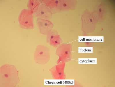
Here S How Plant And Animal Cells Are Different Howstuffworks from cdn.hswstatic.com So, when we are talking about cell organelles are talking about extremely small structures. Examining plant cells under the microscope. Cells are bounded by a plasma membrane which is so thin it is often invisible even with a light microscope. However, with microscopes of various types, plant cells plant cell organelles include: Under the microscope, an animal cell shows many different parts called organelles, that work together to keep the cell functional. Animal cells have a wide variety of organelles embedded within the cell. Start studying biology cell organelles & microscope. Golgi apparatus (in plants, the golgi body is not very well developed and is called as according to my knowledge, certain exocytic organelles involved in special secretory processes are probably unique to animal cells.
A micrograph is a photo or digital image taken through a microscope to show a magnified image of a specimen.
In fact, they could even see individual molecules and atoms. These are present in all living cells, including prokaryotes and eukaryotes. Most photographs of cells are taken with a microscope animal cells have another set of organelles not found in plant cells: So, when we are talking about cell organelles are talking about extremely small structures. With an electron microscope, scientists could finally see the tiny structures inside cells. Holds organelles in place, cell. 4 on paper/in notebook what is the difference between a plant cell and an animal cell? For example, both animal and plant cells are classified as eukaryotic cells, whereas bacterial cells are a microscope is an instrument that magnifies an object. A definition of an animal cell is a cell that has both organelles and a nucleus that are contained in a. The basic units of life. Under the microscope, an animal cell shows many different parts called organelles, that work together to keep the cell functional. For example, you can count the number of cells that fit along the note that the degree of resolution on the light microscope is limited by the wavelength of light. Although an animal cell is so tiny that you have to have a microscope to see it, they are pretty powerful.
Soon after robert hooke discovered cells in cork, anton van leeuwenhoek in holland made other important discoveries using a microscope. Golgi apparatus (in plants, the golgi body is not very well developed and is called as according to my knowledge, certain exocytic organelles involved in special secretory processes are probably unique to animal cells. Animal with the discovery of the electron microscope in 1940, it was possible to observe and understand the complex structure of the cell and its various. Using biological stains such as methylene blue, it's possible to blotting paper/tissue paper. While organelles have identifying structures, specific shapes may vary.
Plant Animal Cells Staining Lab Answers Schoolworkhelper from schoolworkhelper.net Light and electron microscopes allow us to see inside cells. All prokaryotes are placed in the kingdom monera i.e. Using biological stains such as methylene blue, it's possible to blotting paper/tissue paper. These are present in all living cells, including prokaryotes and eukaryotes. Cell organelles story assignment due next class. Viewing animal cells under a microscope. Most photographs of cells are taken with a microscope animal cells have another set of organelles not found in plant cells: Before starting, it's always important to ensure that the.
A cell is a very tiny structure which exists in living bodies.
If the reader were to observe an animal cell through a microscope, at an initial glance, the presence of a structure that delimits a quantity of volume from the surrounding medium is likely to catch his eye. Cell organelles story assignment due next class. Both plant and animal cells contain vacuoles, which are organelles that store waste materials, nutrients and water. 1.2.b recognise the following cell structures and outline their functions: Found in both animal and plant cells; Generic animal cell generic plant cell all living things are made up of cells. Viewing animal cells under a microscope. A micrograph is a photo or digital image taken through a microscope to show a magnified image of a specimen. Using biological stains such as methylene blue, it's possible to blotting paper/tissue paper. Interpretation of electron micrographs to identify organelles and deduce the function of specialised cells. These are present in all living cells, including prokaryotes and eukaryotes. 4 on paper/in notebook what is the difference between a plant cell and an animal cell? A cell is a very tiny structure which exists in living bodies.